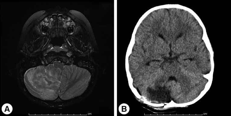
Fig. 1.
Pre- and postoperative imaging studies of the brain. (A) Axial T2-weighted MRI showing a 3.0×4.1×2.5 cm ill-defined infiltrating heterogeneous enhancing mass mainly occupying the right inferior cerebellar hemisphere with perilesional edema causing right tonsillar herniation and mild obstructive hydrocephalus. (B) Follow-up axial CT scan of the brain, 5 days post-operation, showing a hypodense lesion with perilesional vasogenic edema at the right cerebellar vermis and right cerebellar hemisphere, postoperative change.

