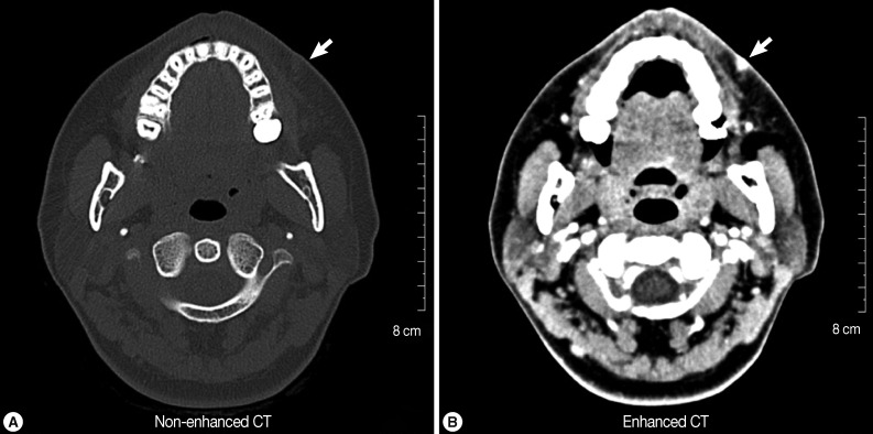
Fig. 1.
Computed tomography (CT) scans (A: non-enhanced; B: enhanced) of the patient showing a small soft tissue mass (arrow) with mild fatty infiltration on the external area of the left nasolabial fold. The lesion migrated slightly to the mucosal side of the upper lip after the CT scan examination and surgery was performed on the lesion.

