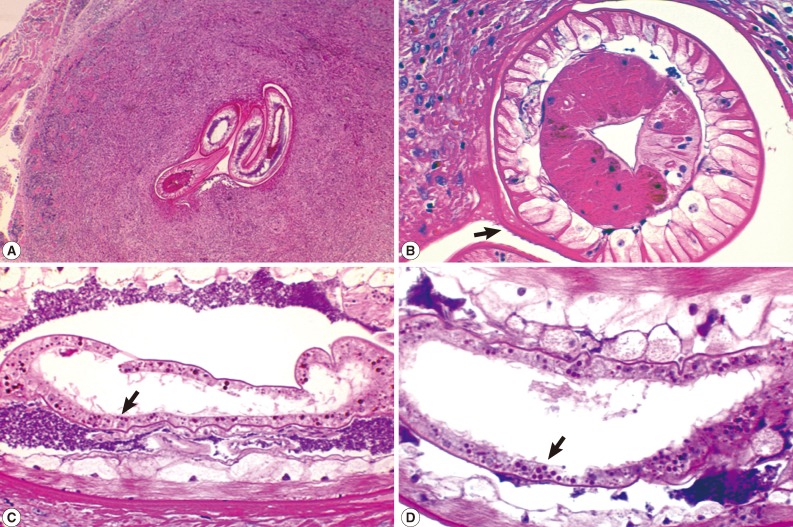
Fig. 2.
Sections of a Gnathostoma spinigerum larva found in the excised mass from the mucosal side of the left nasolabial fold. (A) Sections of the larva showing its anterior (right 2 sections) and posterior (left 1 section) parts (×40). (B) A cross section of an anterior part of the larva showing the cuticle (see cuticular spines; arrow), hypodermis, muscles, lateral cords, and intestine (×200). (C) Another section showing the morphology of the intestine and intestinal cells (arrow). There are approximately 25 intestinal cells, each with 3-7 nuclei (×200). (D) A close-up view of the intestine and intestinal cells; each cell has multiple (3-7) nuclei (arrow, ×250).

