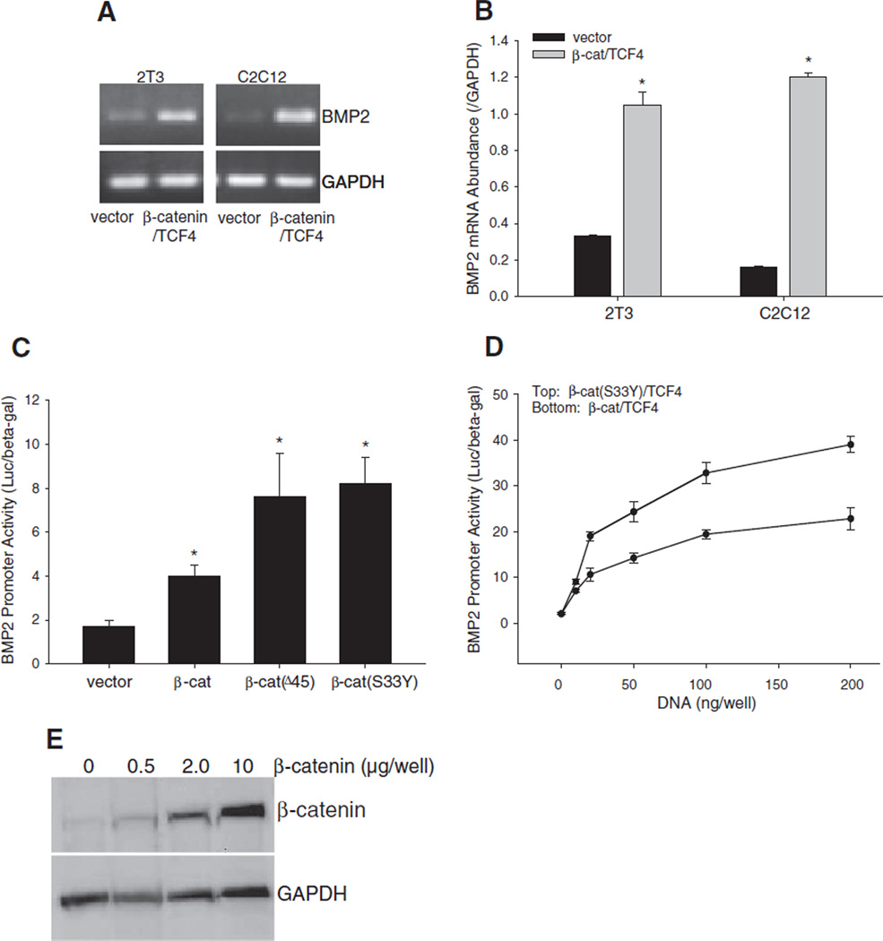
Fig. 4.
Effects of overexpression of β-catenin on BMP2 expression. (A and B) Effects of β-catenin/TCF4 on BMP2 mRNA expression. (A) BMP2 PCR. C2C12 and 2T3 cells were transfected with expression vectors for β-catenin and TCF4 at a dose of 0.5 µg DNA/well in 6-well plates for 24 h. BMP2 mRNA levels in the cell lysates were determined by RT-PCR using mouse BMP2 primers, with GAPDH normalization. (B) Quantitative real time PCR of BMP2. C2C12 and 2T3 cells were transfected with β-catenin/TCF4 as described above. BMP2 mRNA concentrations in the cell lysates were quantitated by real time PCR using TaqMan mouse BMP2 probe, and normalized to GAPDH. *: P<0.05 β-catenin/TCF4 vs. vector (n=4). (C and D) Effects of β-catenin/TCF4 on BMP2 promoter activity. C2C12 cells, which were transfected with −2712/+165-Luc reporter, were co-transfected with expression vectors for wild-type or mutant β-catenin at a dose of 0.2 µg DNA/well in 24-well plates (C) or co-transfected with β-catenin/TCF4 or β-catenin(S33Y)/TCF4 at a dose range of 0.05–0.2 µg DNA/well in 24-well plates (D) for 36 h. Relative luciferase activity in the cell lysates was measured and normalized by β-gal activity. *: P<0.05 β-catenin or its mutants vs. vector (n=6). (E) β-Catenin protein level correlates with amount of plasmid vector transfected. C2C12 cells in 6-well plates were transfected with β-catenin expression vector at varying doses (0.5, 2.0 and 10 µg DNA/well). After 36 h, cells were harvested and β-catenin protein levels in the respective cell lysates were determined by Western blot using anti-β-catenin antibody. GAPDH was used as an internal control.

