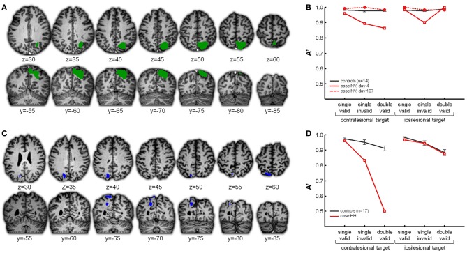
Figure 2.
(A,B) Case NV. (A) In green the right middle IPS lesion in case NV, affecting the horizontal segment of IPS with extension into the superior parietal lobule. (B) NV's accuracy (expressed as A′) obtained in the different conditions of the hybrid spatial cueing paradigm (red), compared to age-matched controls (black). NV was tested on two instances. On day 4 (full red line) the lesion was as visualized in (A), on day 107 the lesion had substantially regressed (dotted red line). For more details see Gillebert et al. (2011). (C,D) Case HH. (C) In blue the left posterior IPS lesion in case HH. (D) HH's performance obtained in the different conditions of the hybrid spatial cueing paradigm (red), compared to age-matched controls (black).

