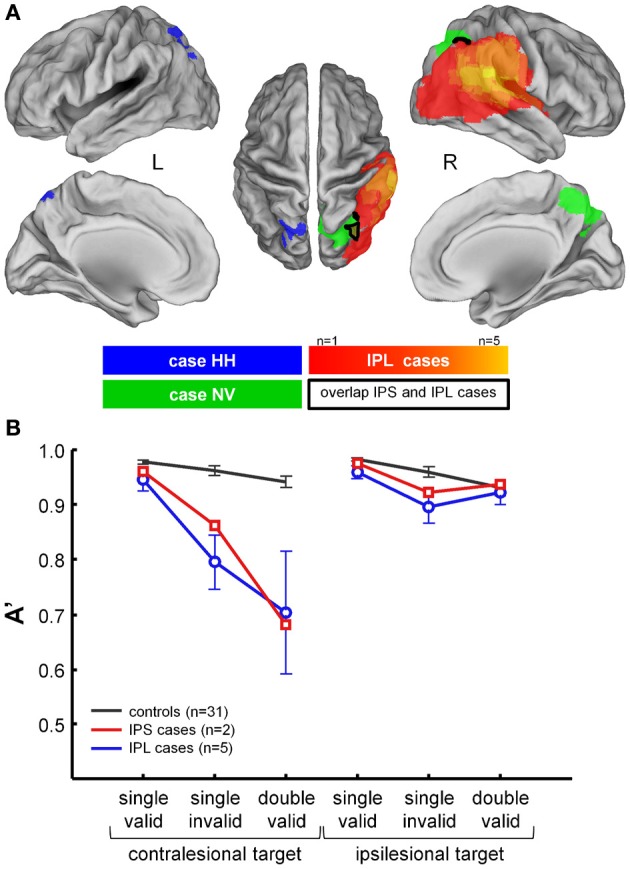
Figure 5.

Comparison between performance in NV and HH during the hybrid spatial cueing paradigm and five cases with classical lesions of right IPL due to ischemia of the territory of the posterior branch of the middle cerebral artery. (A) Projection of the lesion in HH (blue), NV (green), and the IPL cases (hot scale). (B) Average performance in HH and NV (red), in the IPL cases (blue), and in controls (black). For further details see Gillebert et al. (2011).
