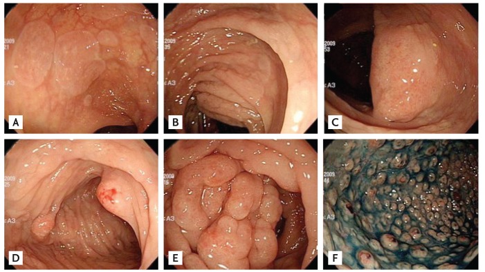
Figure 1.
Initial colonoscopy findings. (A) Proximal colon mucosa shows circular, flat elevated lesions and (B) a very long flat elevated lesion. (C) A mass lesion is seen with (D) a large polypoid lesion resembling Cap polyposis, and (E) a large multigranular mass-like lesion throughout the whole colon. (F) The sigmoid colon and rectum mucosa shows multiple small polypoid lesions resembling familial adenomatous polyposis.

