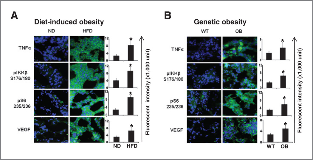
Figure 2.
Increased TNFα/IKKβ/ mTOR/VEGF in mammary tumor of genetic and dietary obesity mice. Representative images of mmunofluorescence staining of mammary tumors with TNFα, VEGF, phosphorylated IKKβ (S176/180) [pIKKβ (S176/180)], and phosphorylated S6 (S240/244) [pS6 (S240/244)] antibodies (right). A, mammary tumor in C57BL/6J female mice that were fed a HFD or normal diet. B, mammary tumor in genetic obese B6.V-Lepob/J (OB) mice that were fed normal chow diet. The relative fluorescence intensity from the images is shown on the right. The data represent the mean ± SD. ND, normal chow diet. *, P < 0.05.

