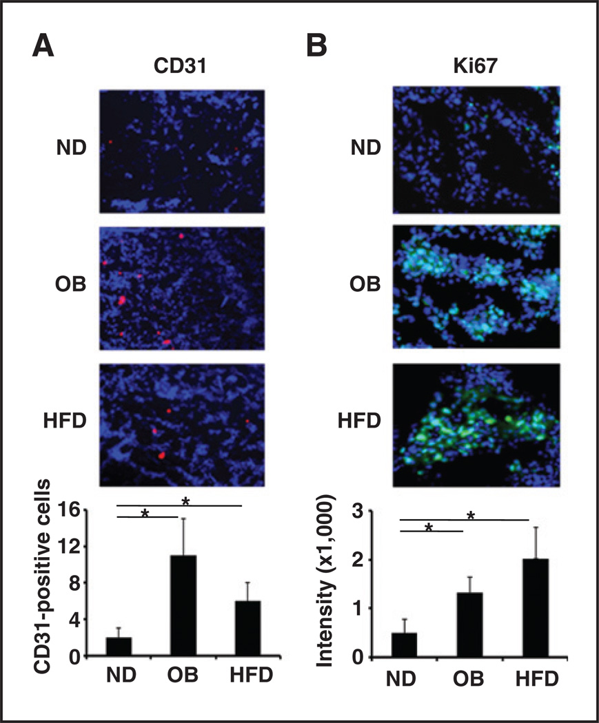
Figure 3.
Increased tumor angiogenesis and tumor cell proliferation in mammary tumors in obese mice. Representative images of immunofluorescence staining of mammary tumor tissues:CD31 antibody as an indicator of tumor vascular density (red; A) and Ki67 antibody as an indicator of tumor cell proliferation (green; B). The relative fluorescence intensity from the images is shown below. The data represent the mean ± SD. ND, normal chow diet. *, P < 0.05.

