
Figure 7.
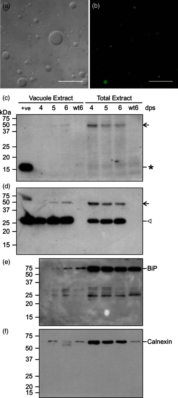
Isolation and analysis of vacuoles from BY-2 cells expressing IL-10-GFP. (a) D.I.C. microscope image of purified vacuoles. (b) GFP fluorescence in purified vacuoles. Scale bars = 30 μm. (c–f) Western blot analysis for IL-10-GFP cell suspension at 4–6 dps. Samples containing 10 μg of protein extracted from purified vacuoles or 10 μg of total soluble protein were immunoblotted with antibodies for: (c) IL-10; (d) GFP; (e) HSC70 (BiP); (f) calnexin. The first lane in panels (c) and (d) (+ve) contains 20 ng of recombinant IL-10 or 20 ng of recombinant GFP, respectively, as positive controls. Asterisk: IL-10, open arrow: IL-10-GFP, open arrowhead: GFP cleavage product. Molecular weight markers, in kilodaltons, are shown on the left; dps, days postsubculturing; wt, wild type.