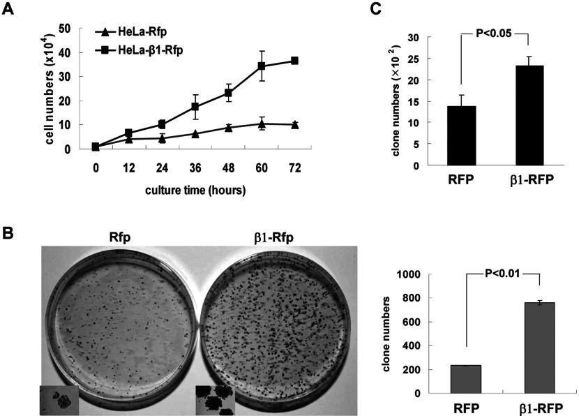
Figure 2. β1 promotes cell proliferation.
(A) Proliferation of HeLa cells stably expressing Rfp and β1–Rfp was determined by cell counting at the indicated times (h) 2h after cells were plated. Data represent an average of three independent experiments. (B) Colonies formed by HeLa cells (500 cells) stably expressing Rfp and β1–Rfp were stained with crystal violet and counted (left panel). Colonies in the small square boxes were documented by a fluorescence microscope (Nikon, ×100). Numbers of colonies are shown in the right panel. Error bars show S.D. (C) Plasmids expressingβ1-Rfp or Rfp were transiently transfected into HepG2 cells and cells were cultured with Gly418 at 0.5 mg/ml for 10 days before staining with crystal violet as described under the Materials and Methods section. It was clear that β1 can promote colony formation by HepG2 cells.

