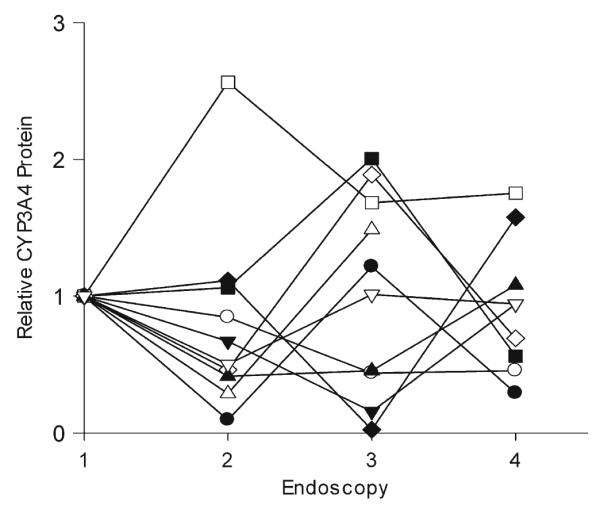
Fig. 4.

CYP3A4 protein expression in intestinal mucosa. Duodenal biopsy tissue was homogenized, and CYP3A4 protein expression was quantified by western blot analysis. The relative amount of CYP3A4, normalized for villin content, at each endoscopy (1–4) is shown for ten subjects (different symbols)
