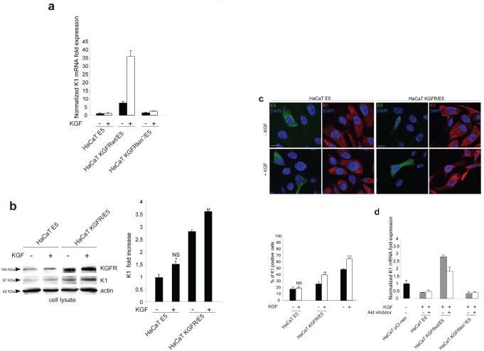
Figure 5. The KGFR counteracting effect on 16E5 requires receptor activation and PI3K/Akt signaling.
(a) HaCaT cells transfected with 16E5 (HaCaT E5) or cotransfected with 16E5 and KGFR (HaCaT KGFRwt/E5) or KGFRY656F/Y657F kinase negative mutant (HaCaT KGFRkin−/E5) were treated with TG as above, serum starved and then stimulated with 20 ng/ml KGF for 24h at 37°C. The K1 mRNA transcript levels were quantitated by real-time relative RT-PCR: a very strong ligand-dependent increase of K1 mRNA expression is observed in HaCaT KGFRwt/E5 cells, although a clear enhancement of K1 levels is detectable also in absence of ligand stimulation, due to a not complete receptor signaling shut down by serum starvation. No significant changes are found in response to KGF treatment in HaCaT E5 cells, as well as in HaCaT KGFRkin−/E5 cells. (b) Western blot analysis on HaCaT E5 and HaCaT KGFR/E5 cells treated with TG and stimulated with KGF as above shows that the band of K1 protein, already increased in HaCaT KGFR/E5 compared to HaCaT E5 cells, is further enhanced in cells stimulated by the ligand. The equal loading was assessed with anti-actin antibody. The densitometric analysis and Student't test were performed and significance levels have been defined as above: *NS vs the corresponding unstimulated HaCaT E5 cells; **p<0,05 vs the corresponding unstimulated HaCaT KGFR/E5 cells. (c) Quantitative immunofluorescence analysis shows a significant ligand-dependent increase of K1 positive cells in KGFR/E5 cells as well as in the surrounding cells that do not express detectable levels of 16E5 protein compared to HaCaT E5 cells. No significant changes are observed in HaCaT E5 cells in response to ligand stimulation. The quantitative analysis was assessed as previously described. Results are expressed as mean values ± standard errors (SE). Student's t test was performed and significance level has been defined as above: *NS vs the corresponding unstimulated HaCaT E5 cells; **p<0,05 vs the corresponding unstimulated HaCaT KGFR/E5 cells; p< 0,001 vs the corresponding unstimulated cells. Bar: 10 μm. (d) HaCaT pCI-neo, HaCaT E5, HaCaT KGFRwt/E5 and HaCaT KGFRkin−/E5 cells were serum starved, treated with TG and then stimulated with KGF in presence or not of the Akt inhibitor. Quantitative real-time RT-PCR shows that the inhibition of Akt reduces the counteracting effect exerted by KGFRwt on K1 down-modulation mediated by 16E5 in HaCaT KGFRwt/ E5 cells while it does not affect K1 levels in HaCaT E5 as well as in HaCaT KGFRkin−/E5 cells.

