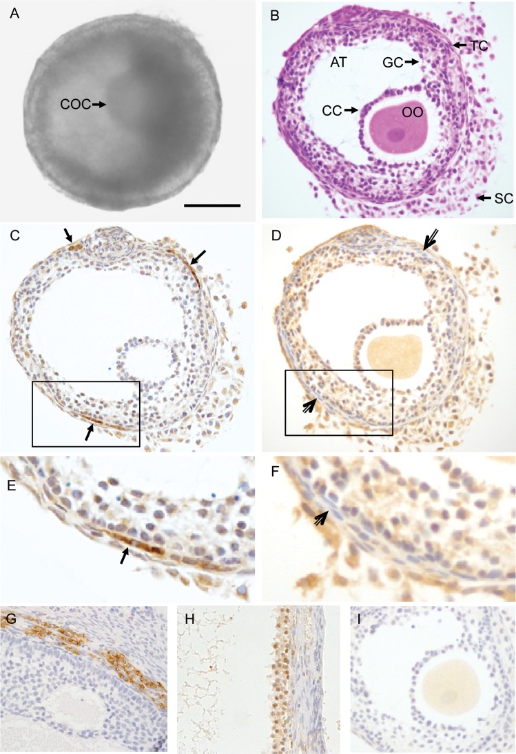
Figure 3.
Histology and immunohistochemical staining of an in vitro-developed macaque follicle. An early antral follicle (A) developed from a primary follicle cultured in FIBRIN was stained with hematoxylin and eosin (B), and immunostained for cytochrome P450 family 17 subfamily A polypeptide 1 (CYP17A1) (C) and cytochrome P450 family 19 subfamily A polypeptide 1 (CYP19A1) (D). The image in the box of C was enlarged in (E), and D was enlarged in (F). Positive control staining for CYP17A1 and CYP19A1 on previously archived macaque ovarian tissue sections is illustrated in (G) and (H), respectively. The non-specific staining associated with the preabsorbed CYP19A1 antibody is illustrated in (I). Arrows, positive staining for CYP17A1 in theca cells. Open arrows, theca cells negative for CYP19A1 staining. Scale bar = 100 µm. COC, cumulus–oocyte complex; OO, oocyte; AT, antrum; CC, cumulus cells; GC, granulosa cells; TC, theca cells; SC, stroma cells.

