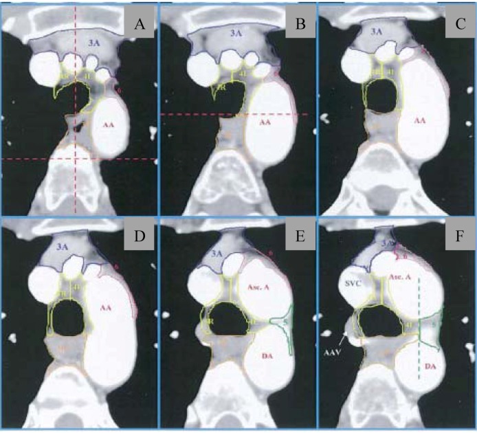
Figure 1.
Thoracic atlas delineating lymph node stations from the University of Michigan (from Chapet et al. [23]). (A–C) Lymph node stations near level of sternal notch; (D) Superior aspect of aortic arch; (E) Level demonstrating posterior limit of 4R and 4L (red dotted line). (F) Level of left brachiocephalic vein, with delineation of 4L/3P and 5. AA = aortic arch; 3A = prevascular nodes; 3P = retrotracheal nodes; 4L/4R = lower paratracheal nodes; 5 = paraaortic nodes; 6 = paraaortic nodes.

