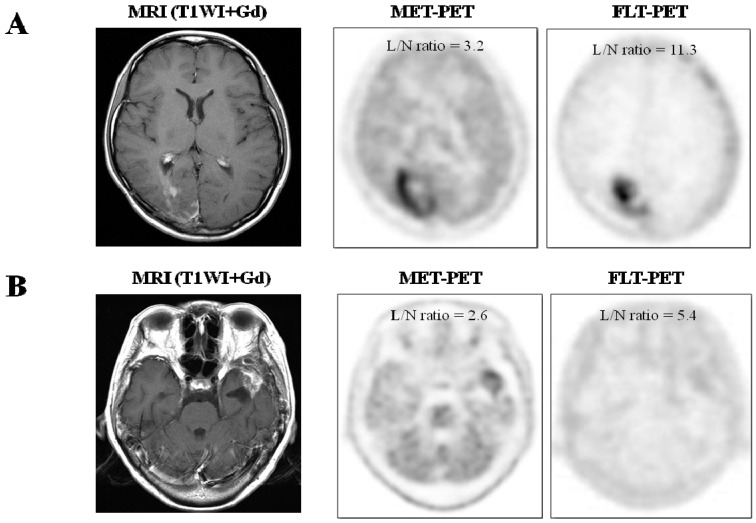
Figure 1.
(A) Imaging of a 22 year-old female (case 9) with glioblastoma, previously treated with tumor resection followed by conventional radiotherapy and temozolomide. T1-weighted contrast enhanced MR image showing irregular enhancement in the right occipital lobe. MET-PET and FLT-PET show intense uptake of tracer in the lesion. Recurrent glioblastoma was pathologically confirmed by surgery; (B) Imaging of 61 year-old female (case 21) with glioblastoma, previously treated with tumor resection followed by conventional radiotherapy and temozolomide. T1-weighted contrast enhanced MR image showing enhanced mass in the left temporal lobe. MET-PET shows mild uptake of tracer and FLT-PET shows faint uptake of tracer in the lesion. Necrosis and gliosis dominant tissue was pathologically confirmed by surgery.

