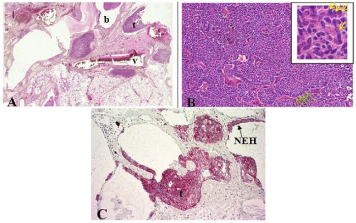
Figure 2.
(A) Cross section of lung specimen with peribronchiolar fibrosis inducing bronchial enlargement. Tumorlet (t) adjacent to bronchus (b) and vascular (v) tissue; (B) High power magnification of tumorlets with neuroendocrine growth pattern, cells are uniform, round with stippled chromatin (yellow arrow); (C) NEH with epithelial proliferation and tumorlets immunopositive for synaptophysin.

