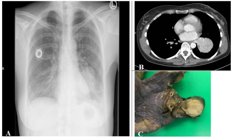
Figure 5.
(A) Corresponding chest radiograph and (B) thorax CT scan (parenchymal window) of a patient with a tissue density/mass (left lower lobe compression) induced by an intrabronchial localized carcinoid tumor demonstrated in the lobe resection tissue (C). The tumor is seen as white to yellow tan intrabronchial tissue mass.

