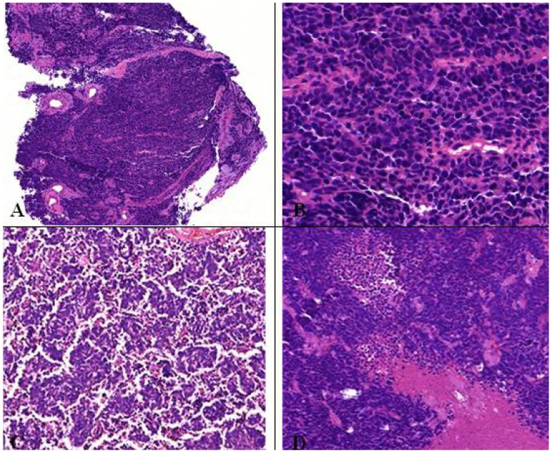
Figure 10.
(A,B) Microscopic feature of small cell lung cancer infiltrate in a bioptical obtained specimen with high mitotic rate. (C,D) Small cell lung cancer in higher magnification with malignant small cells with scant cytoplasm and defined cell borders, fine granular nuclear chromatin, absent or inconspicuous nucleoli, focal or extensive necrosis.

