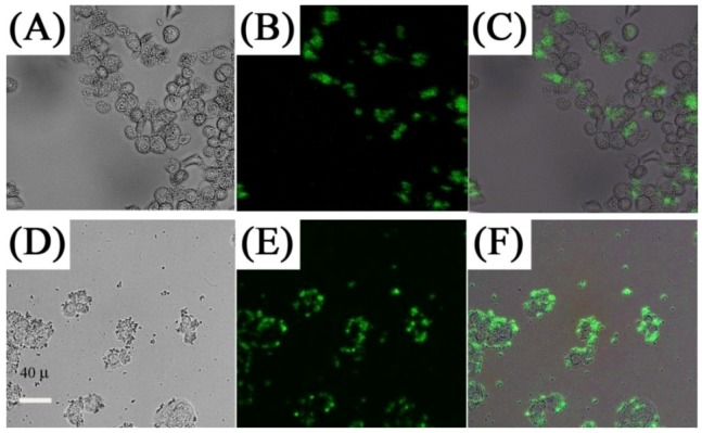
Figure 11.
In vitro imaging using folic acid modified PEI/NaYF4 nanoparticles The top row shows the bright field (A), confocal (B), and superimposed images of live human ovarian carcinoma cells (OVCAR3) (C) and the bottom (E, F) human colonic adenocarcinoma cells (HT29) ([71], reproduced by permission of Elsevier).

