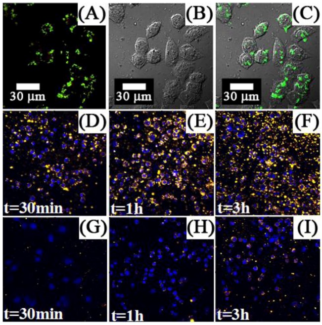
Figure 12.
First row: luminescence (A), bright-field (B) and overlap of both images (C), of KB cells cultured with UCNPs-Ad/β-CD ([123], reproduced by permission of the Royal Society of Chemistry). Second and third rows: Fluorescence imaging of MB49 cells cultured with: carbonized glucose-coated UCNPs (D), (E) and (F); silica-coated UCNPs (G), (H) and (I) ([120], reproduced by permission of the Institute of Physics).

