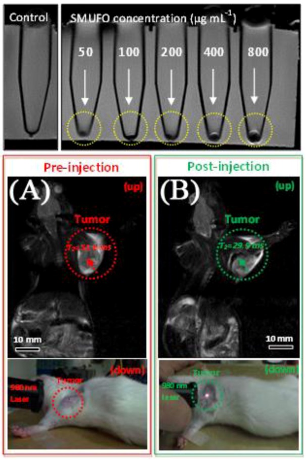
Figure 17.

Top: sensitivity of in vitro MRI of SMUFO treated MCF-7 cells at different concentrations. Bottom: in vivo MR and upconversion fluorescence bimodal imaging of Walker 256 tumor using SMUFO. In vivo whole body pre-injection (A) and post-injection (B) MRI of mouse bearing a Walker 256 tumor. Pre-injection image (A bottom) and post-injection image (B bottom) of the upconversion fluorescence imaging of tumor in vivo using NIR laser ([115,125], reproduced by permission of Wiley).
