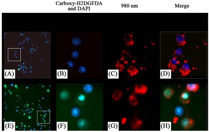
Figure 20.
Detection of oxidative stress in live cells using MB49-PSA cells treated with NaYF4 nanoparticles (A–D) or ZnPc-loaded NaYF4 nanoparticles (E–H) followed by laser activation to induce oxidative stress. Insets in (A) and (E) show the region that has been enlarged in (B–D) and (F–H) respectively. Presence of reduced oxygen species are shown in green, (green) nuclei in blue, and the nanoparticles are shown by their red fluorescence color ([76], reproduced by permission of Elsevier).

