Figure 1.
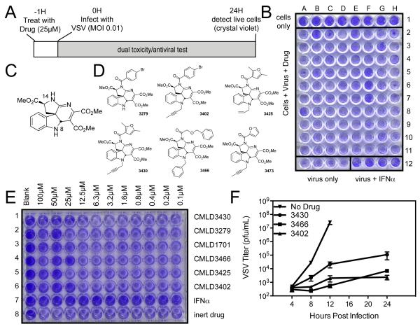
Screen for antiviral compounds using VSV cytotoxicity (A) Schematic of screen. (B) Sample plate from screen: HeLa cells seeded in all wells and treated with library compounds in rows 2-11 and IFNa control in row 12, columns E-H. Infected with VSV MOI 0.1 in rows 2-12 for 24 hours, then fixed and stained with crystal violet. (C) Basic structure of hit scaffold from preliminary screen. (D) Structures of active, indoline alkaloid-type CMLD-BU compounds identified in the screen. (E) HeLa cells were treated with dilution curve of CMLD-BU compounds for 1 hour before infection with wtVSV, then fixed and stained as in screen. (F) HeLa cells were treated with 50μM CMLD-BU compounds for 1 hour before infection with wtVSV at MOI 0.01. Virus was collected at specified timepoints and titer determined by plaque assay.

