
Fig. 5.
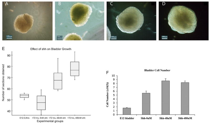
Shh regulates bladder mesenchyme proliferation. Light microscopy (10 ×) images of (A) E12.5 intact bladder prior to organ culture. (B) Intact bladder after 72 h of incubation in serum-free DMEM without Shh. The bladders incubated without Shh were smaller than (C) the bladders incubated with 48 nM of Shh and (D) the bladders incubated with 480 nM of Shh. (E) Box plot representation of the number of sections obtained from intact bladders at harvest and after 72 h of incubation in 0, 4.8, and 480 nM of Shh. The box is the graphical representation of the distribution of data between the first and third quartiles and the line within the box represents the median bladder size. The “whiskers” represent 1.5 times the interquartile range of the pertinent data set. There were no outliers. (F) Cell counts (× 105) of intact bladders at harvest (E12.5) and after 72 h of incubation with increasing concentrations of Shh. After 72 h, the cell counts of intact bladders incubated without Shh nearly tripled relative to preculture levels. Intact bladders cultured with Shh had a greater number of cells than bladders cultured without Shh although there was no difference in the cell counts of intact bladders incubated in 48 nM versus those incubated in 480 nM of Shh.