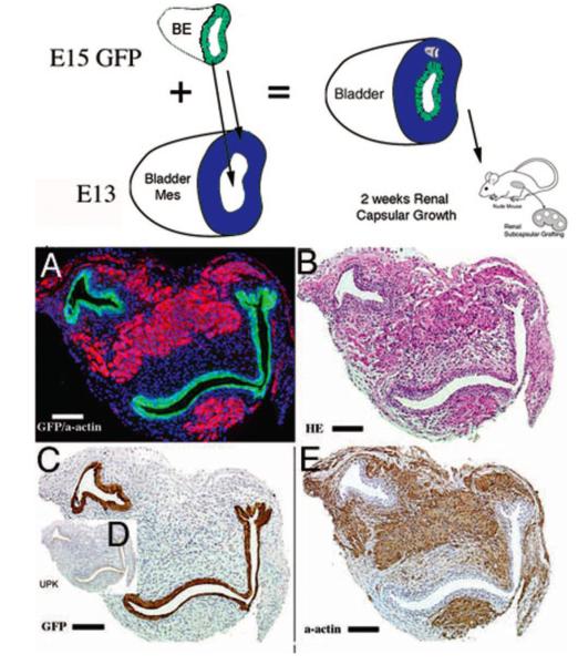
Figure 3. Epithelium patterns bladder smooth muscle.
Schematic and results of urothelial recombination with bladder mesenchyme in the orthotopic location. Histologic serial sections: (A) Color triple florescent stain, GFP is green, alpha-smooth muscle actin is pink and Hoescht dye is blue representing the zone of smooth muscle inhibition or submucosa; (B) H&E dbff hematoxylin and eosin; immunohistochemistry: (C) GFP = green fluorescent protein; (D) UPK= uroplakin and (E) α-actin = smooth muscle alpha-actin. (magnification bar = 100 μm). Reproduced with permission Cao et al., Pediatric Research 2008. [6]

