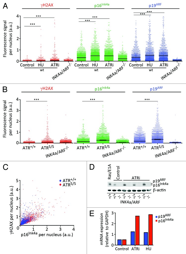
Figure 2. Activation of INK4a/ARF in response to RS. (A) HTM-mediated quantification of the nuclear levels of γH2AX, p16INK4a and p19ARF in wt MEF that had been exposed for 5 d with HU (0.5 mM) and ATRi (1 μM). INK4a/ARF−/− MEF were included as a negative control. (B) HTM-mediated quantification of the nuclear levels of γH2AX, p16INK4a and p19ARF in ATR+/+ and ATRS/S MEF. INK4a/ARF−/− MEF were included as a negative control. (C) 2D plot illustrating the relationship between nuclear γH2AX and p16INK4a levels found on ATR+/+ and ATRS/S MEF. (D) Western blot analysis of p16INK4a and p19ARF in wt MEF that had been exposed to ATRi for 5 d (0.05, 0.1 and 0.5 μM). MEF that had been infected with a retrovirus expressing RasV12 and E1A oncogenes were used as a positive control of INK4a/ARF activation. INK4a/ARF−/− MEF were included as a negative control. (E) qRT-PCR analysis of the mRNA levels of p16INK4a and p19ARF in wt MEF that were grown in the presence of ATRi (2 μM) or HU (0.5 mM) for six passages. mRNA levels were normalized to the expression of GAPDH in each case. Data are representative of three independent analyses. In (A–C) at least 2,000 nuclei were quantified per condition. ***p < 0.001.

An official website of the United States government
Here's how you know
Official websites use .gov
A
.gov website belongs to an official
government organization in the United States.
Secure .gov websites use HTTPS
A lock (
) or https:// means you've safely
connected to the .gov website. Share sensitive
information only on official, secure websites.
