
Figure 3.
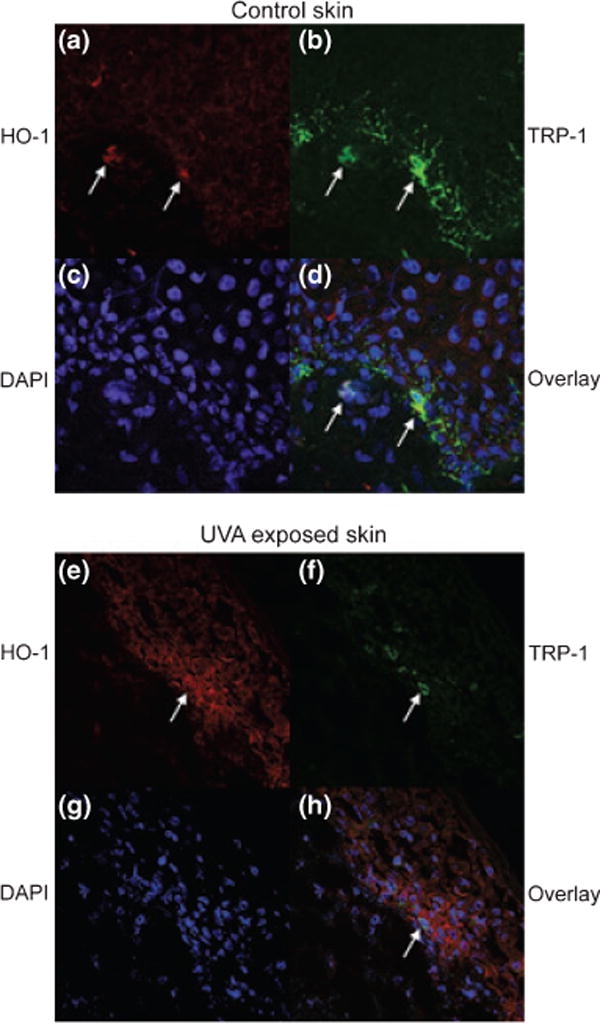
Confocal microscopy of cultured human skin treated with UVA. Skin before and after 1.5 J/cm2 UVA was immunostained with (a, e) anti-HO-1 antibody and (b, f) anti-TRP-1 antibody, and (c, g) DAPI. In (d, h) overlays are shown. (a–d) untreated skin. (e–h) 24 h after UVA treatment. Arrows point to the same cells under (a)–(d) and under (e)–(h).