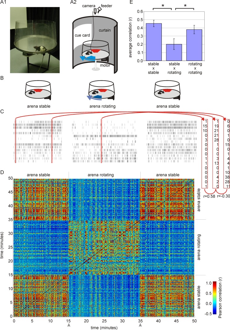
Figure 1. Hippocampal ensemble discharge during the stable and rotating conditions.
(A) A photograph of a rat on the arena (A1) and a schematic drawing of the apparatus during the rotating condition (A2). A rat was placed on a circular arena that was surrounded by a black curtain with a white cue card. The arena rotated slowly in the rotating condition and was otherwise stable. The rat was reinforced to avoid two unmarked shock zones. A room frame shock zone (red) was defined relative to room landmarks and did not rotate. An arena frame shock zone (blue) was defined relative to arena landmarks and rotated together with the arena as indicated by the blue arrow. (B) Schematics of the experimental protocol. Each hippocampal ensemble was recorded during one session of rotating condition, flanked by two sessions of the stable condition. (C) Raster plots of activity of an ensemble of 15 cells during the stable and rotating conditions in the same environment. For each 10-s interval, the ensemble activity was characterized by a spike-count vector (red rectangles). The similarity of ensemble activity during any two intervals was assessed by computing the Pearson correlation between the corresponding ensemble vectors. (D) The correlation matrix shows that the correlation of ensemble activity for each pair of 10-s intervals recorded during the stable condition tends to be high. Similarly, the intervals recorded during the rotating condition tend to have highly correlated activity. Intervals during rotation were often dissimilar to the intervals during the stable condition, as is indicated by blue pixels. (E) Average of the correlation between ensemble activity during different intervals in the stable and rotating conditions (data from all recordings). Correlations are high when activity in the same conditions is compared and lower when activity is compared between the stationary and rotating conditions.

