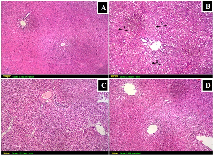
Figure 4. Histopathological findings followed by grading of liver damage.
(x40) (A) control: normal histological structure of portal area and surrounding hepatocytes; (B) CCl4: loss of architecture with severe ballooning degeneration (arrow1), necrosis (arrow2) and fatty changes (arrow3) accompanied with fibrosis; (C) BCA+CCl4: much less damage than in CCl4 gp (D) BCA alone: similar to control gp.

