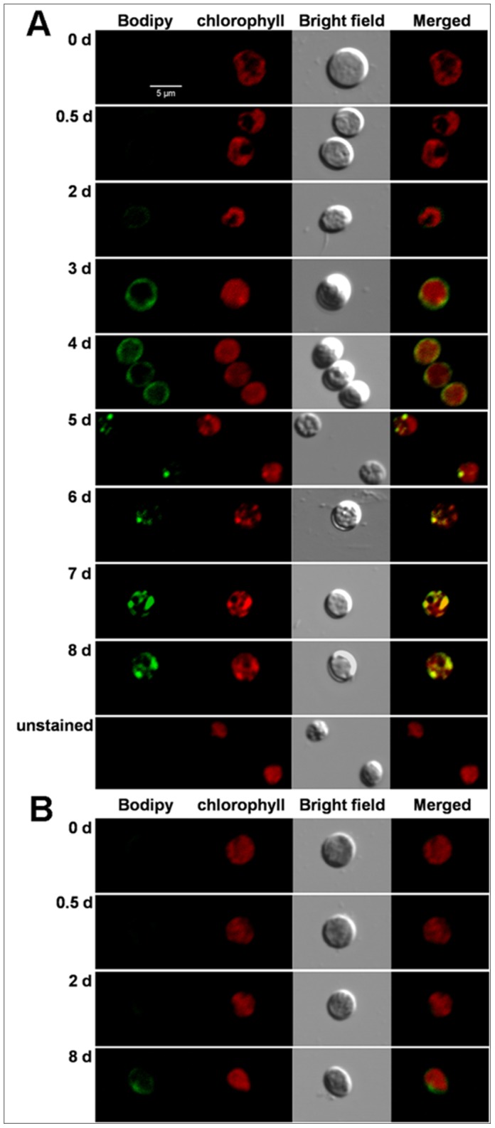
Figure 2. Representative confocal laser scanning micrographs of C. sorokiniana C3 labeled in vivo with Bodipy 505/515.

Bodipy 505/515 (green) was excited with an argon laser (488 nm) and detected at 505–515 nm. Chl autofluorescence (red) was detected simultaneously at 650–700 nm. A, the stained cells resuspended in N− medium for 0 d, 0.5 d, 2 d-8 d, and the unstained cells resuspended in N− medium for 8 d; B, the stained cells resuspended in N+ medium for 0 d, 0.5 d, 2 d and 8 d. The size of the scale bar is shown directly in the image.
