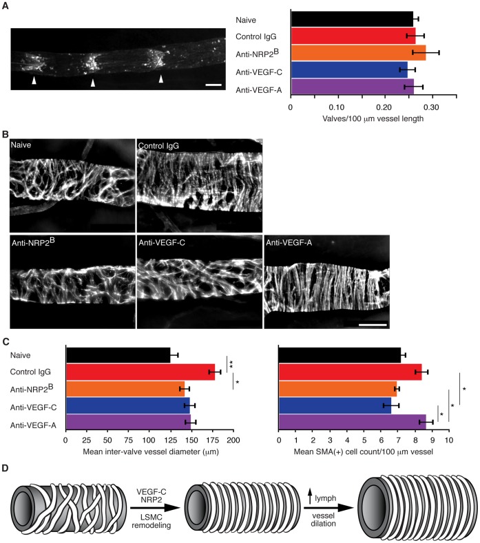
Figure 7. VEGF-C signaling promotes structural remodeling of established lymphatic vessels in metastatic disease model.
(A) Representative epi-fluorescence image (left) of an Ing to Ax lymphatic vessel labeled via intradermal injection of FITC-Lectin. Right, mean valve density ± SEM per C6 tail xenograft tumor and treatment condition: naïve, 0.259±0.013; control IgG, 0.264±0.020; anti-NRP2B, 0.286±0.030; anti-VEGF-C (VC1.12), 0.247±0.018; anti-VEGF-A, 0.260±0.021 valves/100 µm vessel length, n = 5 animals per group. (B) Representative micrographs of αSMA-positive LSMCs along the Ing to Ax vessel. (C) Left, mean Ing to Ax lymph vessel diameter ± SEM measured mid-point between valves: naïve, 125.0±10.5; control IgG, 178.3±8.1; anti-NRP2B, 142.1±6.9; anti-VEGF-C (VC1.12), 148.2±7.5; anti-VEGF-A, 149.4±7.6 µm, n = 14, 12, 13, 12, 14 animals per group, respectively. Right, mean LSMC density ± SEM estimated between lymph valves along the Ing to Ax vessel per condition: naïve, 7.20±0.34; control IgG, 8.41±0.43; anti-NRP2B, 6.95±0.18; anti-VEGF-C (VC1.12), 6.63±0.51; anti-VEGF-A, 8.71±0.44 LSMC/100 µm vessel, n = 7, 5, 6, 8, 7 animals per group, respectively. (D) Model of tumor-associated structural remodeling of distal lymphatics. Scale bar equals 100 µm in A and B. For indicated treatment conditions, tumor-bearing animals were dosed weekly for three weeks starting two days after xenograft implantation.

