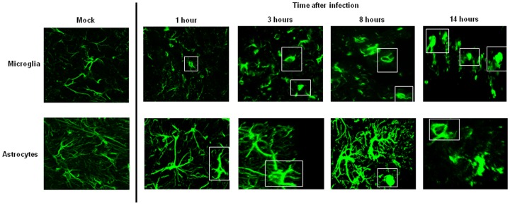
Figure 7. Activation of the local immune system in the brain upon pneumococcal infection.
Immunofluorescent staining of Iba-1 as marker for microglia (A) and GFAP as marker for astrocytes (B) in brain of mock treated mouse and during all the time points of pneumococcal infection. Total magnification 630X. The white rectangles delineate the activated microglia and astrocytes. Brains from 3 mice for each time point were analyzed, and for each mouse 3 brain sections were used for the confocal imaging analysis. Each time point is representative the situation observed in each mouse that was analyzed.

