
Figure 3. Snai1-3 protein expression suggests post-translational regulation.
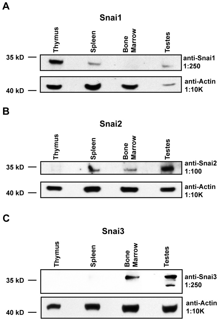
Whole cell lysates were generated from total thymus, spleen, bone marrow, and testes. Samples were subjected to immunoblot analysis as described in the Materials. (A–C) Samples were probed with primary antibodies specific for Snai1 (A), Snai2 (B), and Snai3 (C). Blots were probed for β-actin to verify equal protein loading. Representative blots are shown but similar results were generated for two independent mice.