
Figure 7. Histological analysis of 4 week old DKO spleen and thymus.
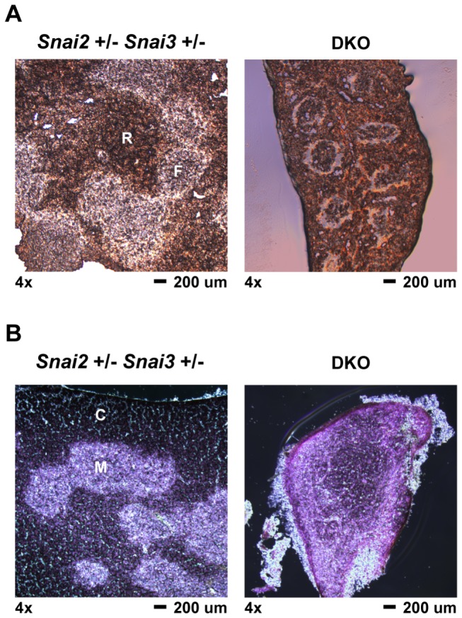
Spleen and thymus were dissected from 4 week old Snai2+/- Snai3+/- and DKO animals. Organs were processed for histological analysis as described in the Materials. Tissue sections were cut to an approximate thickness of 10 µm. Representative images are shown for all genotypes and tissues assayed. (A) Splenic sections were left unstained and viewed by brightfield microscopy. 4x magnification of spleen sections from the Snai2+/- Snai3+/- and DKO. F = lymphoid follicle; R = red pulp. The DKO spleen is reduced in size compared to the Snai2+/- Snai3+/- spleen. (B) Thymus sections were stained with hematoxylin and eosin to differentiate between thymic cortex (darker stain in Snai2+/- Snai3+/-) and thymic medulla (lighter stain in Snai2+/- Snai3+/-): Snai2+/- Snai3+/- (left panel) and DKO (right panel). C = cortex; M = medulla. The DKO thymus is reduced in size compared to the Snai2+/- Snai3+/- thymus.