
Figure 8. Histological analysis of six month DKO spleen and thymus tissues.
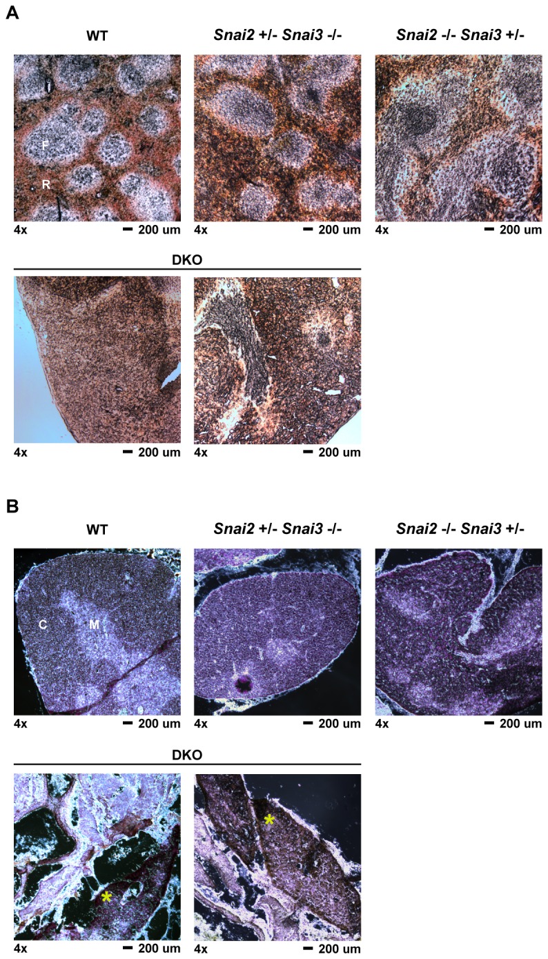
Spleen and thymus tissues were harvested from six month old mice and processed as described in the Materials. Tissue sections were cut to an approximate thickness of 10 µm. All images were captured at 4x magnification. Representative images are shown for all genotypes and tissues assayed with two different sections shown for the DKO samples. (A) Spleen samples from the animals as marked. Sections were kept unstained and viewed via light microscopy for easier assessment of follicular versus red pulp areas of the spleen. F = lymphoid follicle; R = red pulp (B) Thymus sections were stained with hematoxylin and eosin to differentiate between thymic cortex (darker stain) and thymic medulla (lighter stain). C = cortex; M = medulla. An * is shown in the DKO sample to highlight the localization of thymus-like epithelial tissue.