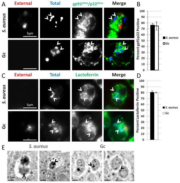
Figure 1. S. aureus and Gc phagosomes fuse with secondary and tertiary granules in primary human PMNs.
(A–D) Adherent, IL-8 treated, primary human PMNs were infected for 1 h with S. aureus or Gc. Extracellular S. aureus and Gc appear red/blue, while intracellular S. aureus and Gc appear blue only. Both S. aureus and Gc infected PMNs were stained with antibodies against the secondary and tertiary granule proteins gp91phox and p22phox (A) or the secondary granule protein lactoferrin (C), which appear green. Arrowheads indicate bacterial phagosomes positive for the granule proteins. The percent of S. aureus and Gc phagosomes positive for gp91phox and p22phox and for lactoferrin are reported in B and D, respectively. (E) Immuno-TEM showing phagosomes positive for lactoferrin that contain S. aureus or Gc. Arrowheads indicate clusters of gold particles accumulated within bacterial phagosomes.

