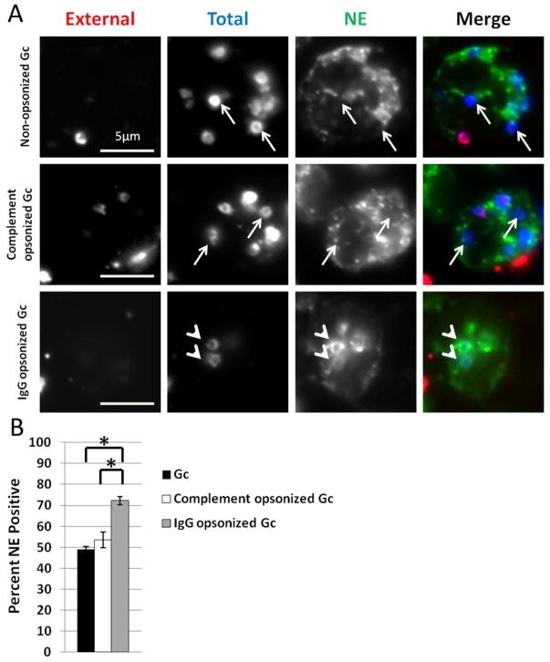
Figure 7. IgG opsonization of Gc increases primary granule fusion with Gc phagosomes.
A. PMNs were infected with non-opsonized, complement opsonized, or IgG opsonized Gc for 1 h, and intracellular and extracellular Gc were discriminated from one another, along with an antibody directed against neutrophil elastase. Extracellular Gc appear red/blue, while intracellular Gc appear blue only, and neutrophil elastase staining appears green. Arrowheads indicate bacterial phagosomes positive for granule proteins, while arrows indicate phagosomes negative for granule proteins. The percent of neutrophil elastase-positive phagosomes containing non-opsonized, complement opsonized, or IgG opsonized Gc is reported in B. Asterisks indicate P < 0.05 by Student’s two-tailed t test.

