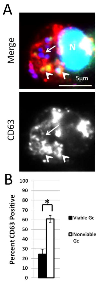
Figure 8. Increased enrichment of the primary granule protein CD63 on phagosomes containing nonviable Gc.

PMNs were infected with DAPI-labeled Gc (blue) for 1 h. Infected PMNs were exposed to soybean lectin (purple) to detect extracellular Gc, then permeabilized and incubated with an antibody against CD63 (red) along with Sytox Green to detect nonviable Gc (green). Viable intracellular Gc appear blue, and nonviable intracellular Gc appear green/blue. A representative infected PMN is shown in A. Arrowheads indicate bacterial phagosomes positive for granule proteins, while arrows indicate phagosomes negative for granule proteins. The percent of viable and nonviable Gc residing in CD63+ phagosomes is reported in B. Asterisks indicate P < 0.05 by Student’s two-tailed t test.
