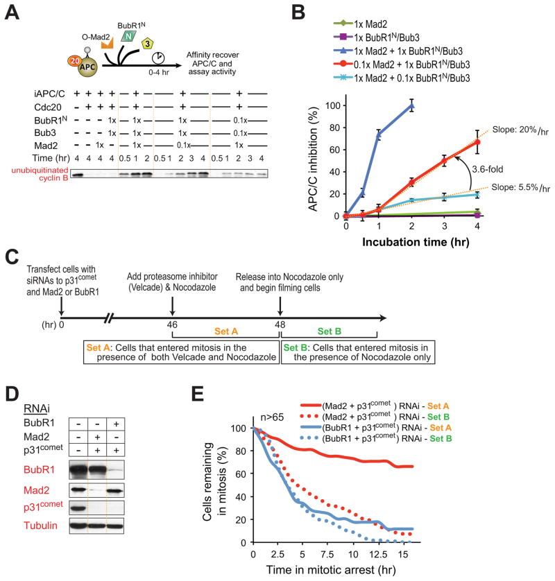
Figure 6. Mad2 catalytically amplifies formation of the final BubR1-APC/CCdc20 inhibitor.
(A) The indicated amounts of Mad2, Bub3, and BubR1N were incubated with preassembled APC/CCdc20 and at various time points assayed for the inhibition of APC/C-mediated ubiquitination of cyclin B1-102. (B) APC/CCdc20 inhibition versus time for the assays denoted in (A). Data represent mean ± SEM (n=3). (C) Schematic of the protocol used to reduce endogenous p31comet and Mad2 or BubR1 levels by siRNA transfection, followed by inhibition and release with the 26S proteasome inhibitor (with 25 nM Velcade) and microtubule inhibitor nocodazole (100 ng/ml), so as to provide a temporal delay in mitotic exit and allow accumulation of mitotic checkpoint inhibitor(s), respectively. (D) Levels of depletion of p31comet, Mad2 or BubR1 by siRNA transfection. (E) Duration of mitosis determined by time-lapse microscopy for cells treated as in b. Filming began after removal of Velcade and release into nocodaozle. Cells counted in Set A entered mitosis in the presence of both nocodazole and proteasome inhibition, whereas in Set B only cells that entered mitosis after removal of the proteasome inhibitor were included. More than 65 cells were analyzed for each condition.

