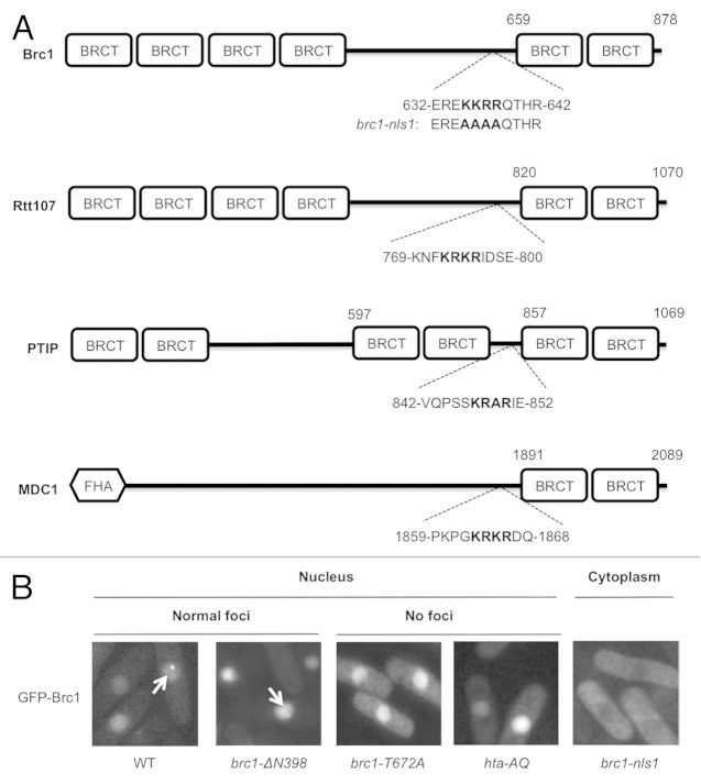
Figure 3. The nuclear localization signal (NLS) of Brc1. (A) Predicted NLS sequences in γH2A-binding proteins. The highest scoring NLS sequence calculated using cNLS Mapper is shown for each protein. The brc1-nls1 mutant allele is also indicated. (B) Live-cell microscopy of wild type, truncated and mutant GFP-Brc1. GFP-Brc1 was expressed from pREP42-GFP-brc1 plasmids. Cells were grown in EMM (Edinburgh minimal media) for 18–20 h at 30°C. Arrows indicate Brc1 foci. Wild type (WT) Brc1 and the N-terminal truncation lacking the N-terminal BRCT domains (brc1-ΔN398) showed normal Brc1 foci formation, whereas the brc1-nls1 mutant fails to localize in the nucleus. The brc1-T672A mutant that cannot bind γH2A localizes in the nucleus but fails to form foci. Wild GFP-Brc1 expressed in hta-AQ also fails to form nuclear foci.

An official website of the United States government
Here's how you know
Official websites use .gov
A
.gov website belongs to an official
government organization in the United States.
Secure .gov websites use HTTPS
A lock (
) or https:// means you've safely
connected to the .gov website. Share sensitive
information only on official, secure websites.
