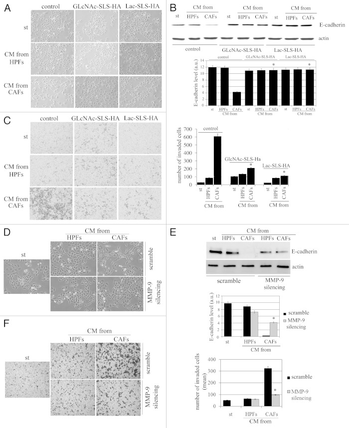
Figure 6. The modulation of MMP-9 expression prevents EMT in PCa cells. (A) Representative images of PCa cells treated for 72 h with CM from HPFs and CAFs containing MMP-9 inhibitor GlcNAc-SLS-HA and Lac-SLS-HA. (B) Immunoblot of E-cadherin expression in the same experimental setting described in (A). (C) Invasion analysis of PCa cells in the presence of MMP-9 inhibitor GlcNAc-SLS-HA and Lac-SLS-HA. Bar graph represents the mean of invaded PCa cells (six fields for sample randomly chosen). (D) MMP-9 was silenced in HPFs and CAFs. Then, CM from HPFs or from CAFs (both control and MMP-9 silenced) were added to PCa cells for 72 h, and cells were photographed. (E) Immunoblot analysis of E-cadherin in PCa cells in the same experimental setting described in (D). Actin immunoblot is used for normalization. (a.u., arbitrary units). (F) Invasion assay of PCa cells treated with CM from control HPFs or CAFs or with CM from silenced-MMP-9 HPFs or CAFs. *p < 0,001 vs. control CAFs.

An official website of the United States government
Here's how you know
Official websites use .gov
A
.gov website belongs to an official
government organization in the United States.
Secure .gov websites use HTTPS
A lock (
) or https:// means you've safely
connected to the .gov website. Share sensitive
information only on official, secure websites.
