Figure 2.

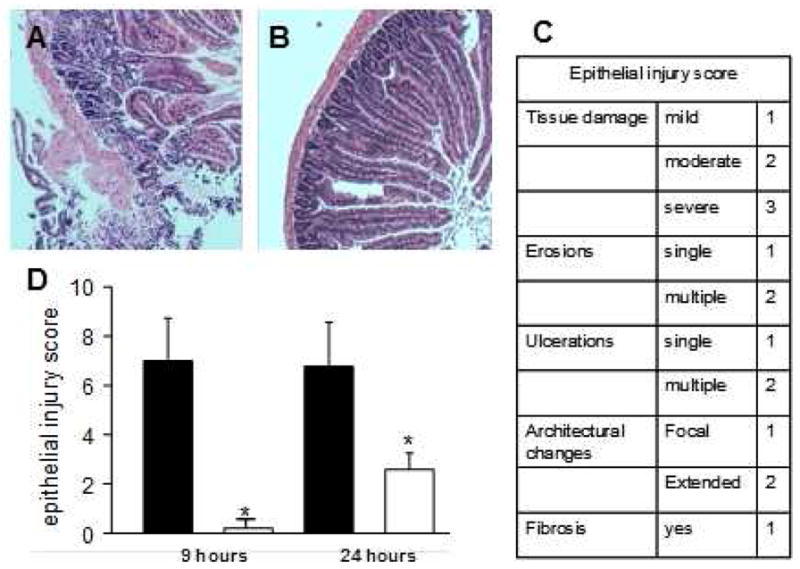
Histological analysis of the donor intestinal grafts. H&E staining of mice donor intestinal grafts preserved in (A) histidine-tryptophan-ketoglutarate (HTK) or in (B) HTK containing 5% PEG 15–20 (HTK/PEG). (C). Epithelial injury score. (D) Evaluation of severity of structural injury of donor intestinal grafts preserved in either HTK (■) or HTK containing 5% PEG 15–20 (□). n=50 donor grafts, *p<0.001.
