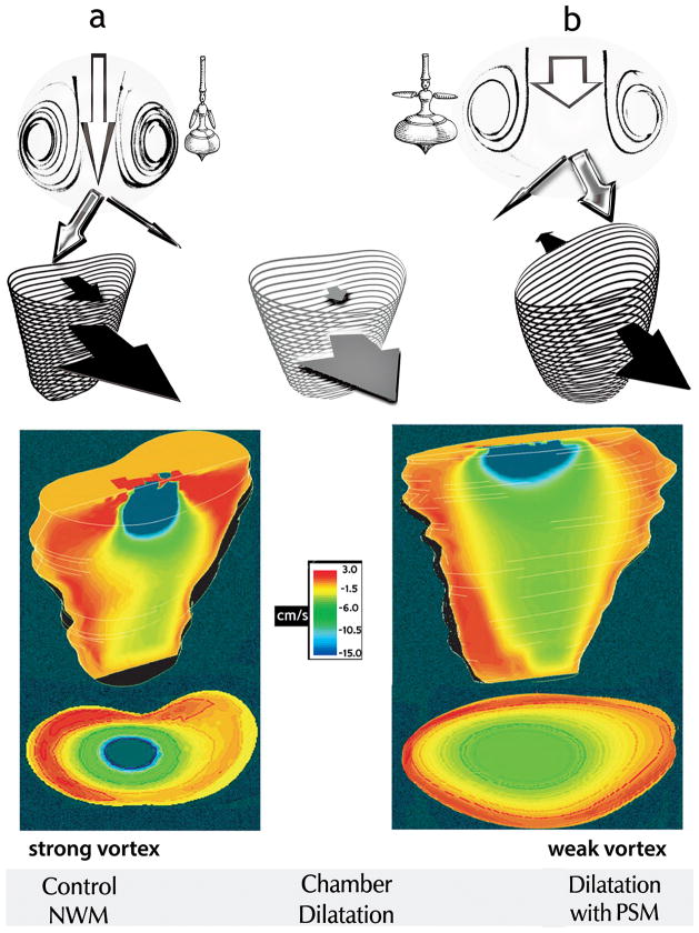
Fig. 4.
Top panels Under control conditions with normal wall motion (NWM), diastolic filling entails anterior-directed motion of both the free RV wall and the septum (black arrows, a). In RV chamber dilatation with paradoxical septal motion (PSM), the septum moves toward the left ventricle (back-pointing black arrow, b). Interposed is the pattern of RV dilatation with normal septal motion (gray arrows). By increasing both intraventricular mass and effective rotation radii, increased chamber size yields smaller recirculating velocities and vortex strength, as the “whirling dervish” tops suggest: with arms extended and wider girth, spinning is slower in b than in a. The stronger vortex ring in the normal-sized chamber (panels a) encroaches more on the central core than the weaker vortex in the enlarged chamber (panels b). Consequently, although instantaneous volumetric inflow rates at control were smaller than with volume overload, after vortex development higher linear core velocities were present at control than with chamber dilatation attendant to volume overload. The width of each white arrow in the top panels is proportional to central core area; the length, to linear inflow velocity.
Bottom panels: Simultaneous frontal-plane and transverse (at a level 2.5 cm below the inflow orifice) sections showing RV color flow maps, close to the end of the E-wave. The instantaneous volumetric inflow velocities were 39 cm3/s in the normal-sized (left) and 71 cm3/s in the dilated (right) chamber, respectively. These Functional Imaging mappings show expediently the higher axial core velocities in the normal and the larger core cross-section in the enlarged chamber. (Color maps reproduced from Pasipoularides et al. [13] by permission of The American Physiological Society.)

