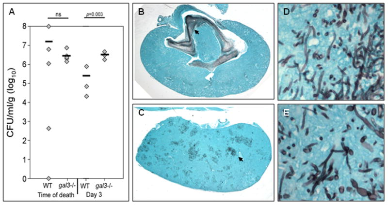
Figure 2. Fungal burden and distribution in kidneys infected with C. albicans.

WT and gal3−/− mice were infected with 1×10⁵ CFU of C. albicans via tail vein injection. (A) Kidney fungal burden of infected WT and gal3−/− mice at time of death and at day 3 post-infection. Comparisons of fungal burdens were made by Mann-Whitney rank-sum test when there were sufficient cases for ranking, or a negative binomial model where group n=3. Bar represents mean CFU. Low magnification view of GMS stained kidney sections of infected WT (B) and gal3−/− mice (C) at time of death. Fungal elements appear black. Black arrows point to dense areas of fungus. High magnification (×600) of GMS stain of kidney of infected WT (D) and gal3−/− (E) mouse demonstrating yeast and hyphal morphologies.
