
Fig. 1.
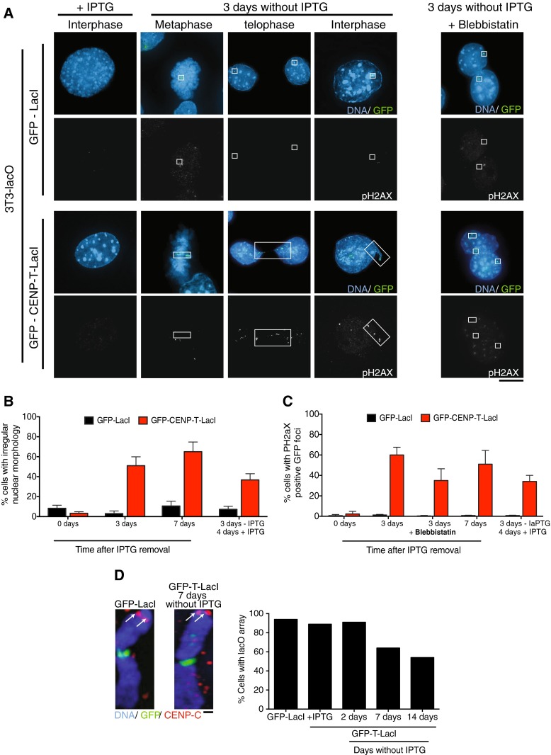
Induction of an ectopic kinetochore causes nuclear abnormalities and DNA damage in 3T3-LacO cells. a Stable cell lines expressing GFP-CENP-T ΔC-LacI or GFP-LacI were generated in 3T3 cells containing a lacO array on chromosome 3. Growth in the presence of 10 mM IPTG prevented LacI–lacO interactions. Representative immunofluorescence images show 3T3-LacO cells after removal of IPTG for 3 days and staining with anti-phospho-H2AX antibodies. Distortion of the GFP-CENP-T-LacI containing chromosome is clearly seen after removal of IPTG, as well as co-localization of DNA damage signal with GFP-CENP-T-LacI. Right panel shows nuclear morphology after removal of IPTG and growth in 30 μM blebbistatin, indicating reduction in nuclear abnormalities after inhibition of cytokinesis. Boxes indicate the location of the GFP signal. Scale bar shows 5 μm. b Quantification of the number of 3T3-lacO cells with irregular nuclear morphology (including nuclear protrusions, multi-lobed nuclei, and separated nuclei connected by chromatin bridges) in the presence of the indicated LacI fusion protein and following removal of IPTG for 3 or 7 days, or removal for 3 days, then re-addition for 4 days. c Quantification of the number of 3T3-lacO cells with phospo-H2AX signals co-localized with GFP foci in immunofluorescence images after growth in the indicated conditions. For b and c; n ≥ 100 cells in three independent experiments. Error bars show standard deviation. d Shows representative images of LacO (green) containing chromosomes that also have an active endogenous centromere in the indicated conditions, as detected by CENP-C immunofluorescence staining in red. Arrows indicate CENP-C-specific staining. Scale bar shows 1 μm. Graph shows the number of 3T3-LacO cells containing the lacO array in the indicated conditions