
Figure 3.
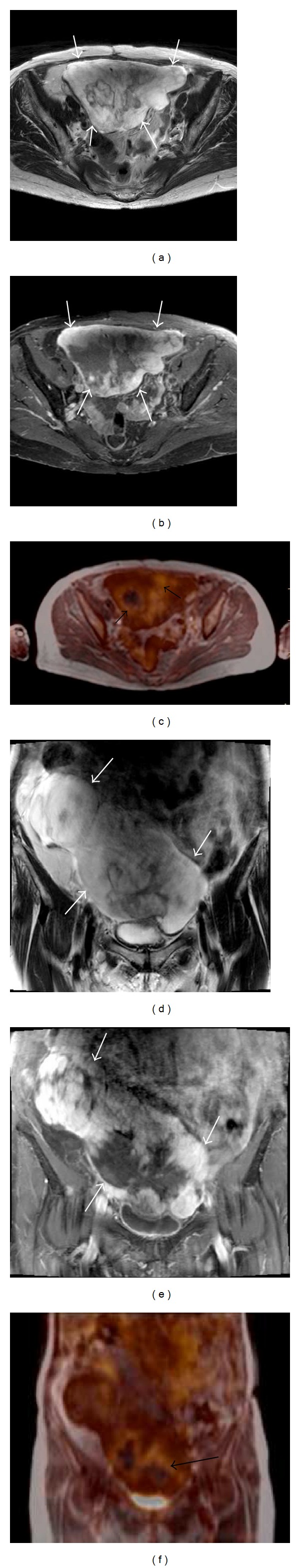
A 68-year-old patient with abdominal liposarcoma. The large tumour (white arrows) is well recognizable on both the T2-weighted images ((a) and (d)) and the contrast-enhanced, fat-saturated T1-weighted images ((b) and (e)). The fused PET/MR images ((c) and (f)) show an inhomogeneous FDG uptake (black arrows).