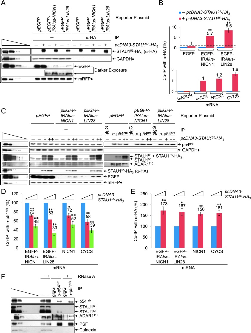
Figure 2.
STAU1 coimmunoprecipitates with 3′ UTR IRAlus in competition with p54nrb. (A) Western blotting of lysates of formaldehyde cross-linked HEK293T cells (1.8 × 10⁷ per 150-mm dish), which had been transiently transfected with 15 μg of pcDNA3-HA (−) or pcDNA3-STAU155-HA3 (+), 5 μg of pmRFP, and 5 μg of the specified pEGFP reporter, before (−) or after immunoprecipitation using anti-HA (Supplemental Table S5). (IP) Immunoprecipitation. (B) Histograms of RT-qPCR analyses of RNA from lysates analyzed in A. (Top) EGFP mRNA levels before immunoprecipitation were normalized to the level of GAPDH mRNA, and EGFP mRNA levels after immunoprecipitation were normalized to the level of LACZ RNA (immunoprecipitated samples were spiked with Escherichia coli RNA prior to RNA extraction). Subsequently, the normalized level of EGFP mRNA after immunoprecipitation is presented as a ratio of its normalized level before immunoprecipitation, and this ratio is defined as 1 for samples transfected with pEGFP + pcDNA3-STAU155-HA3 (Supplemental Fig. S2A). (Bottom) As in the top panel but analyzing cellular mRNAs, where the ratio of c-JUN mRNA after immunoprecipitation/before immunoprecipitation is defined as 1 (Supplemental Fig. S2B). (C–E) HEK293T cells (1.8 × 10⁷ per 150-mm dish) were transiently transfected with 25 μg of pcDNA3-HA (−) or 15 μg (+) or 25 μg (++) of pcDNA3-STAU155-HA3, 5 μg of the designated pEGFP reporter, and 5 μg of pmRFP. Cells were then formaldehyde cross-linked, and half was immunoprecipitated using anti-p54nrb, while the other half was immunoprecipitated using anti-HA. (C) Western blotting of lysates before or after immunoprecipitation using anti-p54nrb or gIgG (Supplemental Table S5). (D) Histograms of RT-qPCR quantitations (Supplemental Fig. S2C) of RNA from samples shown in C, where the level of each mRNA was normalized to the level of GAPDH mRNA and LACZ RNA, respectively, before and after anti-p54nrb immunoprecipitation. Normalized values are provided as after immunoprecipitation/before immunoprecipitation, and the ratio in pcDNA3-HA-transfected cells is defined as 100. (E) Histograms of RT-qPCR quantitations (Supplemental Fig. S2D) are as in D except that each mRNA was normalized to the level of GAPDH mRNA before anti-HA immunoprecipitation or LACZ RNA after anti-HA immunoprecipitation. (F) Western blotting of lysates of HEK293T cells (1.8 × 10⁷ per 150-mm dish), which were generated in the presence (+) or absence (−) of RNase A before or after immunoprecipitation, essentially as in C. All results are representative of three independently performed experiments. (*) P < 0.05; (**) P < 0.01; n ≥ 3.

