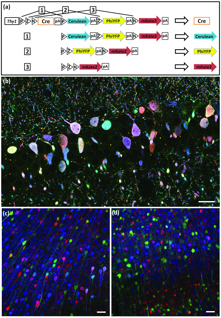
Figure 3. Autobow.
(a) In Autobow, a self-excising Cre recombinase cDNA is placed in the first (default) position. Once expressed, Cre can lead to expression of any of the 3 following XFPs (outcomes 1–3), but is itself excised in the process, terminating recombination.
(b) Labeling of hippocampal neurons by Autobow founder 3. The 20 large neurons (diameter <5µm) in this section are labeled in 20 distinct colors. Antibody amplified Cerulean, PhiYFP and mKate2 are in blue, green and red, respectively.
(c,d) Cortical neurons of an Autobow mouse line show similar color palette in the second (c) and sixth (d) generations. Antibody amplified Cerulean, PhiYFP and mKate2 are in blue, green and red, respectively. Bars are 50µm.

