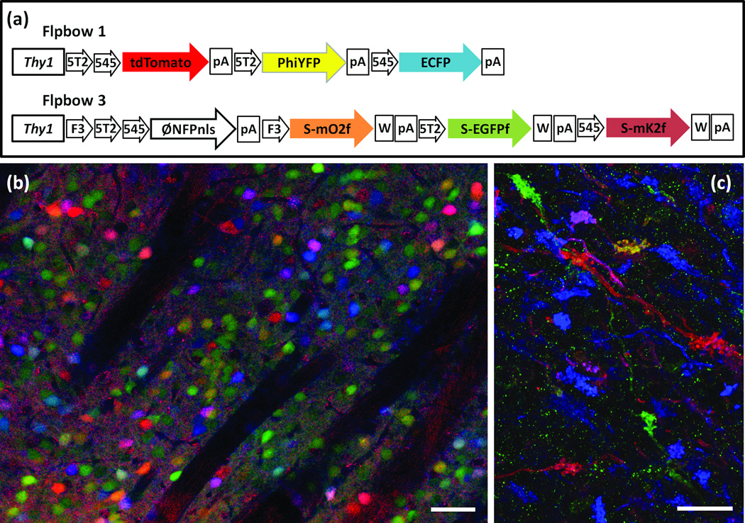
Figure 4. Flpbow.
(a,b) Flpbow transgenes incorporate Frt sites, which are substrates for Flp recombinase, instead of Lox sites, which are substrates for Cre recombinase. In Flpbow 1 (a), the Lox sites of Brainbow 1.0 were replaced by incompatible Frt sites Frt5T2 and Frt545. In Flpbow 3 (b), the Lox sites of Brainbow 3.2 were replaced by incompatible Frt sites F3, Frt5T2 and Frt545. In addition, XFPs in this construct were fused to SUMO-Star, providing an epitope tag.
(c) Labeling of neurons in caudate putamen of Flpbow 1; Wnt-flp double transgenic. Neurons are labeled by at least nine colors (red, orange, yellow, yellow-green, green, cyan, blue, purple, pink). Native fluorescence of ECFP, tdTomato and antibody amplified PhiYFP are in blue, red and green, respectively.
(d) Labeling of mossy fibers in Flpbow3; Wnt-flp double transgenic. Fluorescence of antibody amplified EGFP, mOrange2 and mKate2 are in blue, red and green, respectively.
Bars are 50µm in c, 20µm in d.

